 Echocardiography
 is a non-invasive
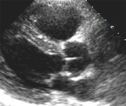
imaging procedure used to evaluate heart chambers and function, heart valves, adjacent heart structures, and blood flow in
and out of the heart.
BCS Heart offers on-site echocardiography
evaluation.
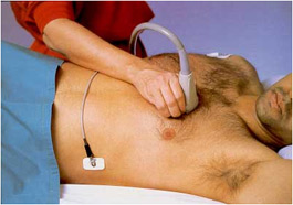
Echocardiography involves the use of sound waves generated and received by a small probe near the surface of the
heart to produce real-time images. The small probe can be positioned on the outside
of the chest (termed transthoracic echocardiography, see image to the right) or inside the esophagus/stomach (termed transesophageal
echocardiography). Transthoracic echocardiography usually is able to provide satisfactory imaging of all areas of
the heart, but occassionally transesophageal imaging may be required.
For more detailed information about the procedure, continue
reading below.
With transthoracic echocardiography, a technician obtains images (see image to right) by placing a small
probe (usually with a lubricated jelly-like material on its tip) at various locations on the skin surfaces of the chest
and abdomen. Rarely, an intravenous (IV) line may be required to administer a medication to enhance image quality or
evaluate for certain shunts (abnormal connections) between heart chambers. The patient is monitored by electrocardiography
throughout the procedure, which typically takes less than 30 minutes. No sedation is routinely required.
With transesophageal echocardiography, a physican, alongside a technician, will obtain images by passing a small probe
into the back of the throat which is then swallowed by a sleeping patient. An intravenous (IV) line is routinely required
to administer sedation medications. The patient is monitored by electrocardiography throughout the procedure, and images
can typically be obtained in under 30 minutes. The patient routinely requires one to two hours of recovery following the
procedure depending on the amount of sedation required. The patient may then be driven home following recovery.
Results of the procedure are typically available the same or following day.